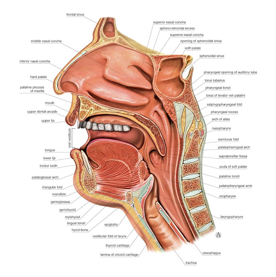
Oral Cavity And Pharynx Photograph by Asklepios Medical Atlas Fine
This e-Anatomy module contains 110 illustrations on the oral cavity, the mouth, the tongue and the salivary glands. These fully annotated anatomical illustrations are presented as a comprehensive atlas of the oral cavity, specially designed for medical students, medicine residents and healthcare professionals. Material and methods
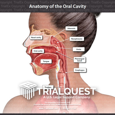
Anatomy of the Oral Cavity TrialExhibits Inc.
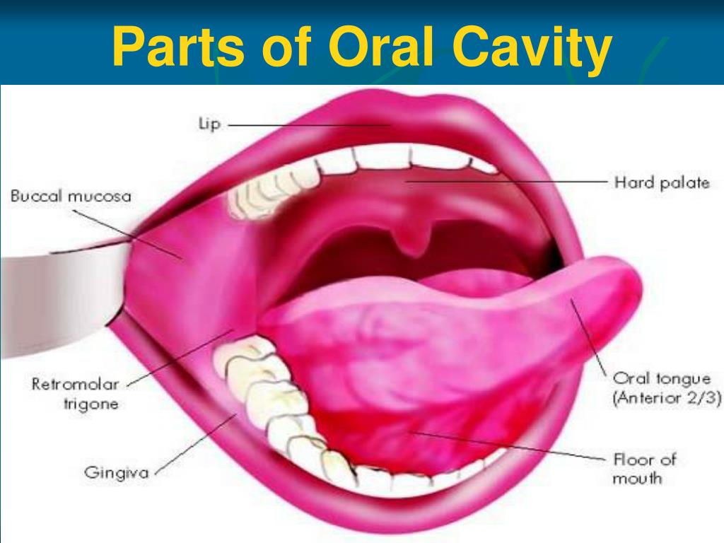
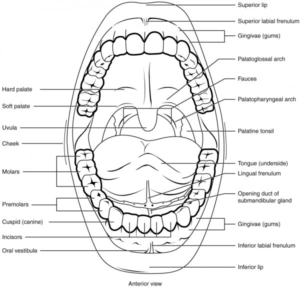
Mouth conditions Where to get help Image: Dental Health Services Victoria The mouth is an oval-shaped cavity inside the skull. The two main functions of the mouth are eating and speaking. Parts of the mouth include the lips, vestibule, mouth cavity, gums, teeth, hard and soft palate, tongue and salivary glands.
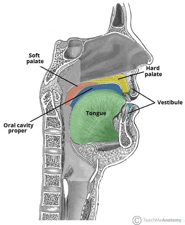
The Oral Cavity Divisions Innervation TeachMeAnatomy
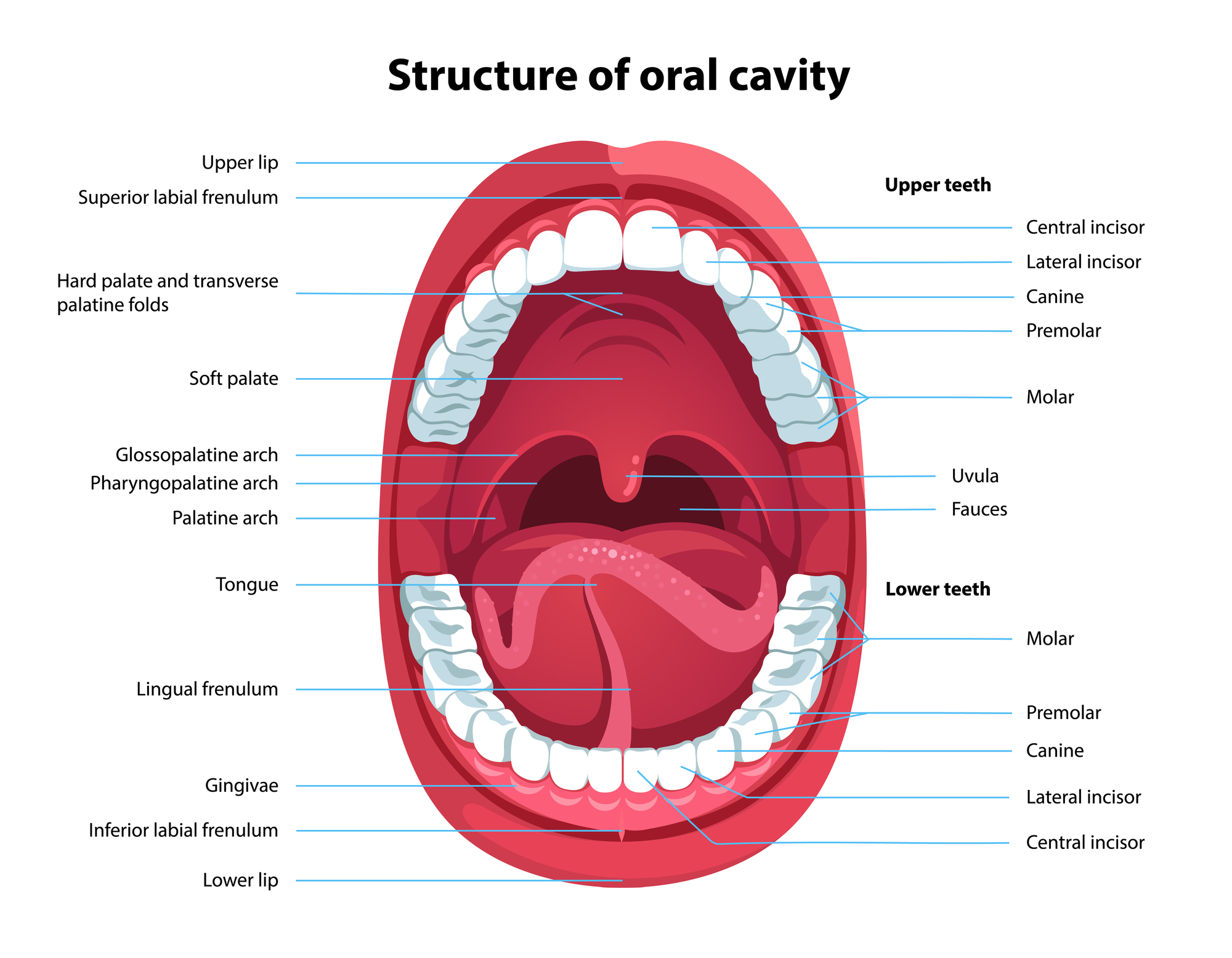
The alveolar bone is covered by specialized oral mucosa known as gingiva, or, more commonly, gums. The remainder of the oral cavity is covered with an oral mucous membrane. Tooth germ formation of the primary or baby teeth begins 7 to 10 weeks in utero. This is followed by enamel and dentin apposition from 4 to 6 weeks in utero.
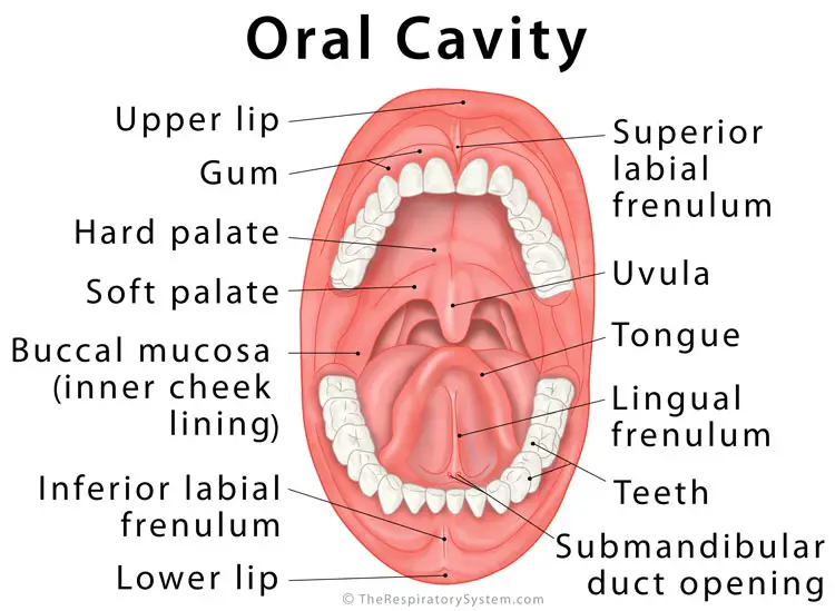
What is the Oral Cavity
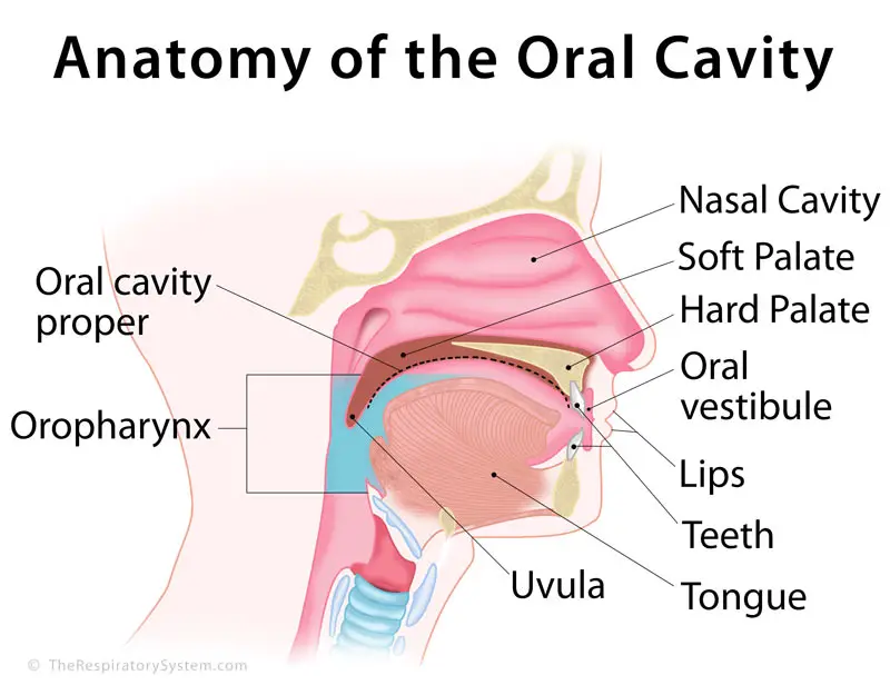
Oral cavity Anatomy of the mouth. Floor of the mouth with lingual frenum and sublingual fold The mouth consists of two regions: the vestibule and the oral cavity proper. The vestibule is the area between the teeth, lips and cheeks. [3]

oral cavity Coding Pinterest
Many parts make up your mouth anatomy. These parts work together harmoniously to help with chewing, speaking and breathing. The outside of your mouth creates a boundary that holds food in place and helps you form sounds and words. It includes your cheeks and lips. The inside of your mouth contains your: Teeth. Gums.
Anatomy of the Oral Cavity YouTube
1/2 Synonyms: none The cavity is separated into anterior and posterior parts by the dental arches (or teeth): the anterior oral vestibule sits anteriorly to the teeth and behind the lips, whilst the oral cavity proper describes the area behind the teeth.

Oral cavity anatomy with educational labeled structure vector illustration
The oral cavity, better known as the mouth, is the start of the alimentary canal. It has three major functions: Digestion - receives food, preparing it for digestion in the stomach and small intestine. Communication - modifies the sound produced in the larynx to create a range of sounds.
Dental Articles and Resources
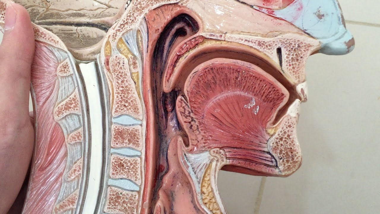
The roof of the oral cavity is formed by the palate, which separates the oral cavity from the nasal cavity above. At the bottom, the mylohyoid muscle forms a muscular diaphragm that supports the tongue and the structures of the floor of the mouth. Finally, the lateral walls of the oral cavity are formed by the cheeks.
PPT Anatomy of Oral Cavity, Pharynx & Oesophagus PowerPoint
The vestibule of the oral cavity refers to the narrow space that lies between the lips and cheeks and the external surface of the teeth and gingivae (gums). The oral cavity proper refers to the region that sits inside the dental arcades. Inferiorly, it's bounded by the muscular floor of the mouth. Superiorly, it's bounded by the palate and.

Structure Of Oral Cavity Human Mouth Anatomy Stock Vector Images and
Subject Dentistry Collection: Oxford Scholarship Online 25.1 Introduction Technically, the oral cavity consists of the vestibule between the lips and cheeks externally and the teeth and alveolar processes internally and the larger oral cavity proper located internal to the dental arches.

AN3 08 Oral Cavity, Oropharynx, Swallowing StudyBlue
. The teeth are hard, calcified structures used for sound articulation as well as biting and masticating food. The oral cavity is separated from the nasal cavity by the palate , which is divided into the soft and the hard palate . The masticatory movements of the jaw are enabled by the muscles of mastication
Oral Cavity Proper
The oral cavity and vestibule are entirely lined by mucous membranes containing numerous small glands that, along with the three pairs of salivary glands, bathe the mouth in fluid, keeping it moist and clear of food and other debris.

Oral Cavity Diagram Unlabeled
Present exceptional content with your unlimited creative subscription. Download Millions of PowerPoint Templates, Graphic Assets, Fonts, Icons & More!

Oral Cavity Anatomy Model Images Galleries With A
References: Neil S. Norton, Ph.D. and Frank H. Netter, MD, Netter's Head and Neck Anatomy for Dentistry, 2nd Edition, Elsevier Saunders, Chapter 13 Oral Cavity, Page 341 to 351. Rudolf Beer, Michael A. Baumann and Andrej M. Kielbassa, Pocket Atlas of Endodontics, 1st Edition, Thieme Flexibook, Chapter 1 Anatomy and Types of Endodontic Pathology, Normal Endodontium, Page 2 and Chapter 5.
What Are the 3 Key Functions of the Teeth? Vancouver Centre for
1 Citations Abstract In this section, the surface structures of the oral cavity, which is necessary to understand the mimetic muscles and floor of the mouth, will be reviewed. Download chapter PDF 3.1 Surface Anatomy of the Oral Cavity Summary
Oral Cavity Anatomy, Functions, and Diseases Medical Library
Dental Care Package at Parramatta Medical and Dental Centre. Get Exclusive Deals With Groupon. Limited Time Offer